The Simple Drawing Of Protein Synthesis offers a clear, visual pathway from DNA to a functional protein. This compact guide uses simple shapes and labeled steps to illustrate transcription and translation, helping learners grasp how genetic information is transformed into everyday biological work.
Key Points
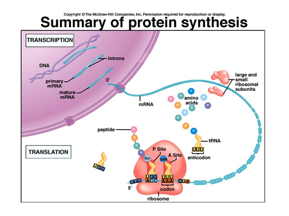
- The diagram separates transcription (DNA to mRNA) from translation (mRNA to amino acid chain) to show two distinct stages.
- Color coding highlights DNA, mRNA, ribosome, tRNA, and the growing polypeptide for quick recognition.
- Directionality is emphasized: the flow from DNA to mRNA and from mRNA to a growing protein chain.
- Codon-anticodon pairing at the ribosome demonstrates how genetic code guides amino acid addition.
- The sketch hints at multiple ribosomes translating one mRNA (polysomes), illustrating efficiency in protein production.
What the drawing shows
The visual starts with DNA in the nucleus, showing the genetic code being copied into messenger RNA. The mRNA then exits the nucleus and encounters a ribosome, where translation begins. Transfer RNA brings amino acids, matching codons on the mRNA with anticodons on tRNA, and the polypeptide chain grows one amino acid at a time. The sketch may indicate a stop signal to mark the end of translation and briefly remind viewers that the resulting protein will fold into its functional shape.
How to read the Simple Drawing Of Protein Synthesis
Look for labeled components: DNA, mRNA, ribosome, tRNA, and the expanding protein strand. Follow the arrows to trace the journey from transcription in the nucleus to translation in the cytoplasm, then imagine how the final folded protein comes from the linear chain produced by the ribosome.
Practical tips for study
Use the drawing as a study aid by recreating it from memory, then labeling each part yourself. Try explaining the process aloud to a peer while pointing to the diagram, and quiz yourself on the order of events—from DNA unwinding to protein release. The simple drawing can serve as a referential backbone for more detailed study notes later.
How do transcription and translation differ in this drawing?
+The drawing places transcription in a nucleus-related area where DNA is used to synthesize mRNA, then shifts to the cytoplasm where the ribosome reads the mRNA to assemble the protein. This separation mirrors the two-step flow of genetic information: DNA → RNA → protein.
What role do codons and anticodons play in the visual?
+Codons on the mRNA determine which amino acids are added next. Anticodons on tRNA pair with those codons, guiding the ribosome to place the correct amino acid in the growing chain. The pairing depicted in the drawing reinforces how the genetic code operates during protein synthesis.
Why is the ribosome a central feature in the diagram?
+The ribosome is the molecular machine that assembles amino acids into a polypeptide by translating the mRNA sequence. In the drawing, it acts as the hub where tRNA brings amino acids and the chain lengthens until a stop signal ends translation.
Can this simple drawing depict different proteins produced by the same gene?
+While the drawing provides a foundational view, in reality, the final protein depends on the sequence of codons read from the mRNA and subsequent folding and post-translational modifications. The visual focuses on the core flow, not the full diversity of protein variants.