Understanding Apmlitude In Xrays: How Subtle Signals Reveal Disease

The term Apmlitude In Xrays refers to the height or strength of light absorption variations that radiologists analyze when reading X-ray images. In modern radiology, Apmlitude In Xrays helps clinicians identify patterns that may indicate disease even when obvious structures appear normal. This article explains what amplitude means in X-ray images, how subtle signals are detected, and what that means for diagnosis and patient care.
Key Points
- Subtle amplitude differences can flag early disease before obvious signs appear.
- Consistent imaging technique is essential for reliable amplitude comparisons.
- Quantitative amplitude metrics accompany radiologist interpretation to improve confidence.
- Artifacts and technical factors can masquerade as amplitude changes and must be ruled out.
- Integrating amplitude insights with clinical data and AI tools can enhance monitoring and outcomes.
How amplitude manifests in X-ray images
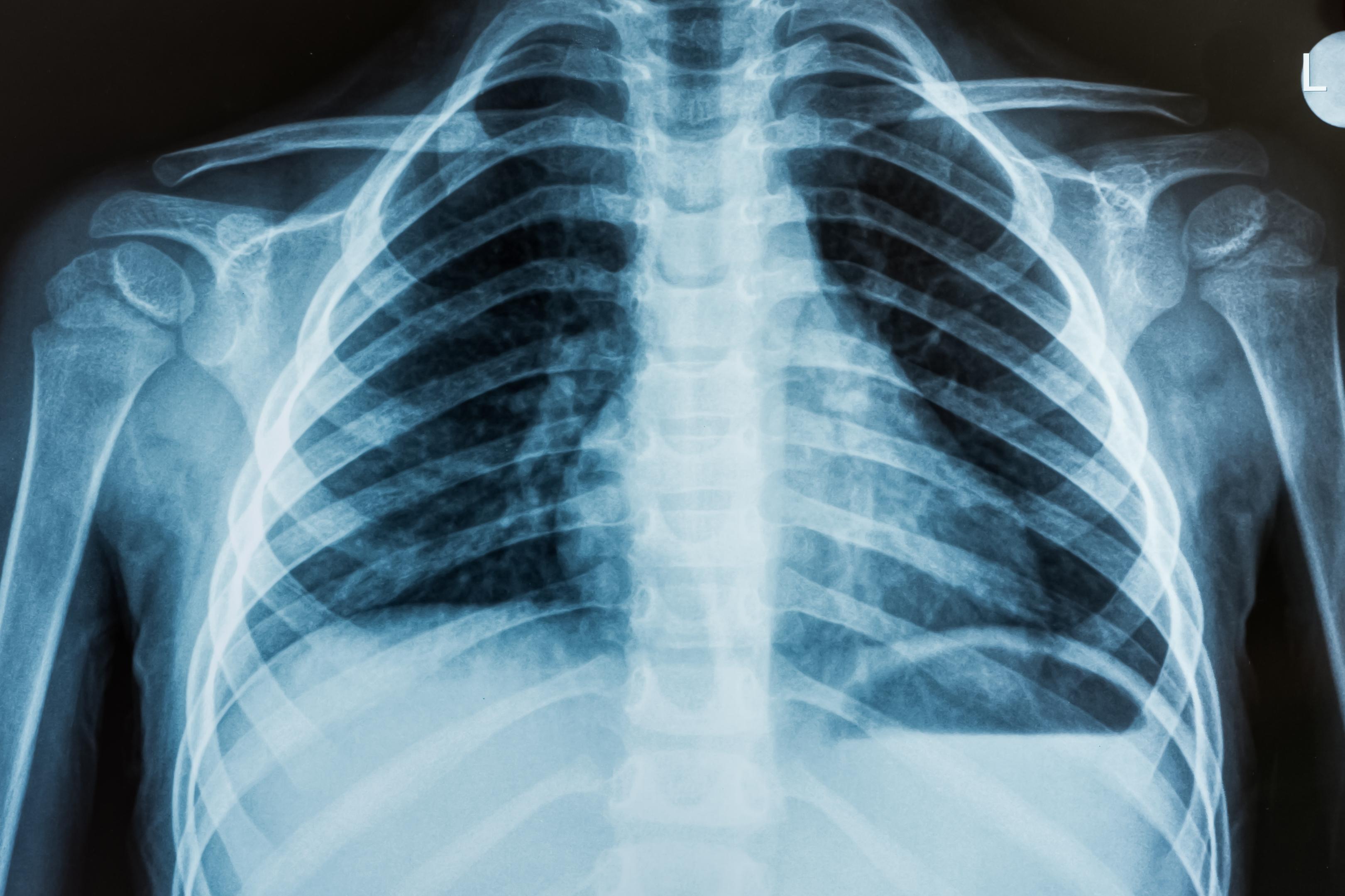
Amplitude in X-ray imaging describes how large the variations are in pixel values that correspond to different tissue densities. Higher amplitude differences can reveal dense structures like bone, while subtle amplitude changes may hint at early disease processes such as infiltrates or fibrosis. Understanding these signals requires good image quality, proper exposure, and awareness of the patient’s clinical context.
Interpreting signals: practical implications for radiology
Clinicians consider amplitude alongside anatomy, symmetry, and distribution. Apmlitude In Xrays is not a standalone diagnosis; it is a clue that guides further testing, follow-up imaging, or additional modalities like computed tomography (CT) or magnetic resonance imaging (MRI). When amplitude changes align with patient symptoms, history, and risk factors, the likelihood of a meaningful finding increases.
Limitations and considerations
Several factors can affect amplitude interpretation. Exposure settings, patient movement, and detector performance can alter pixel values, potentially mimicking or masking disease signals. Recognizing these limits helps clinicians decide when a repeat image or alternative imaging is warranted, reducing false positives and unnecessary interventions.
Towards a more nuanced diagnosis
As imaging advances, amplitude analysis is increasingly complemented by quantitative tools and artificial intelligence. These approaches can quantify amplitude patterns, compare them over time, and integrate with clinical data to support faster, more accurate decision-making.
What does Apmlitude In Xrays measure in simple terms?
+It describes the magnitude of pixel value changes in an X-ray image that reflect how much tissue density attenuates the X-ray beam. Larger amplitude differences often correspond to distinct structures, while subtle changes can indicate early pathology when interpreted with context.
  <div class="faq-item">
    <div class="faq-question">
      <h3>Can small amplitude changes indicate disease even if the image looks normal?</h3>
      <span class="faq-toggle">+</span>
    </div>
    <div class="faq-answer">
      <p>Yes. Small amplitude variations can precede visible findings or reflect early inflammatory, infectious, or fibrotic processes. They are most meaningful when correlated with symptoms, history, and, if needed, follow-up imaging or alternative modalities.</p>
    </div>
  </div>

  <div class="faq-item">
    <div class="faq-question">
      <h3>How do clinicians differentiate true amplitude signals from artifacts?</h3>
      <span class="faq-toggle">+</span>
    </div>
    <div class="faq-answer">
      <p>Clinicians assess exposure settings, patient positioning, and image quality; they may compare with prior studies, repeat the study if necessary, and use additional imaging or processing techniques to confirm whether a signal is pathology-related or an artifact.</p>
    </div>
  </div>

  <div class="faq-item">
    <div class="faq-question">
      <h3>Is amplitude analysis affected by the imaging hardware?</h3>
      <span class="faq-toggle">+</span>
    </div>
    <div class="faq-answer">
      <p>Yes. Detector sensitivity, calibration, and processing algorithms influence amplitude readings. Standardized protocols and regular equipment maintenance help ensure reliable interpretation across exams and institutions.</p>
    </div>
  </div>

  <div class="faq-item">
    <div class="faq-question">
      <h3>What future developments could enhance amplitude-based diagnosis?</h3>
      <span class="faq-toggle">+</span>
    </div>
    <div class="faq-answer">
      <p>Advances in quantitative radiomics, AI-driven pattern recognition, and longitudinal amplitude tracking will enable more precise, reproducible assessments. Combining amplitude data with patient-specific risk factors and other imaging modalities holds promise for earlier and more accurate disease detection.</p>
    </div>
  </div>
</div>